Bone Cancer Patients and Others Receiving 3D-Printed Implants at UH
February 18, 2024
UH Clinical Update | February 2024
Innovations in Orthopaedics | Winter 2024
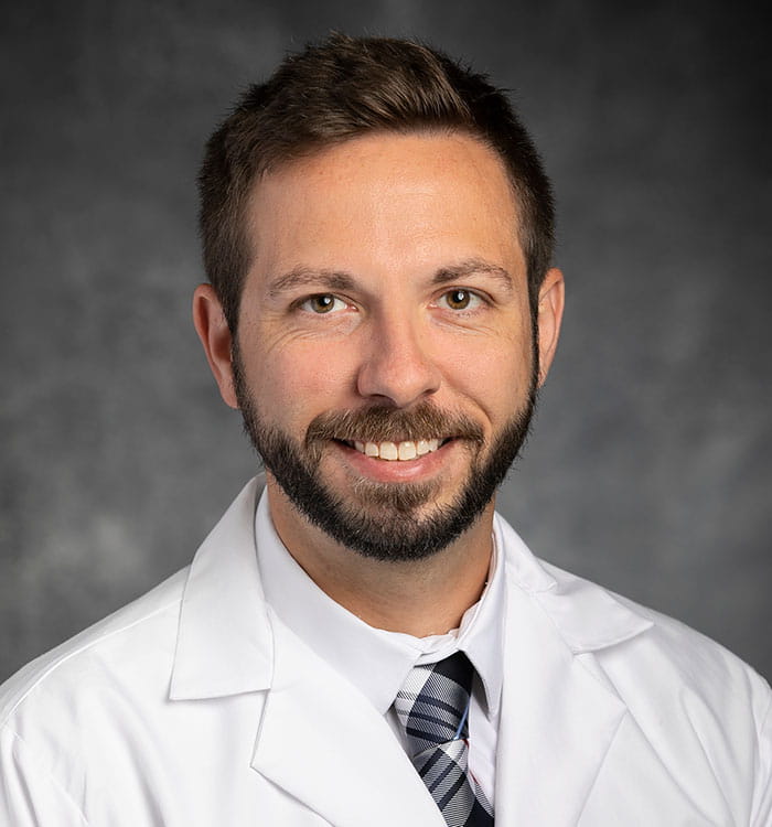
 Brandon Jonard, MD
Every patient undergoing surgery for a cancerous tumor of the bone is different in important ways. But the off-the-shelf implants orthopedic surgeons typically use to restore structure to the affected limb or joint – not so much. When an appropriate implant to meet a patient’s specific needs isn’t available, the consequences can be devastating.
“Sometimes there is no other option for them besides leaving them without a reconstruction,” says University Hospitals orthopedic oncology specialist Brandon Jonard, MD. “We sometimes call that a flail leg.”
A Better Way
Fortunately, Dr. Jonard is giving patients better options. He’s one of a select few orthopedic oncology specialists around the country using 3D printing technology to meticulously design and produce both “jigs” to guide more precise bone cuts during surgery, as well as the implant itself.
 3D Custom Implants.
“It really gives us the advantage of having something that we can utilize to give them function back, when there is no other option off the shelf in terms of implants,” he says.
"We are fortunate to have Dr. Jonard as part of the Department of Orthopaedic Surgery,” says James E. Voos, MD, Professor and Chair, Department of Orthopaedic Surgery, Jack and Mary Herrick Distinguished Chair in Orthopaedics and Sports Medicine and Head Team Physician, Cleveland Browns. “He provides a level of compassion and expertise that comforts patients often facing a challenging diagnosis. Dr. Jonard and his team are using proven and the most cutting-edge treatments to provide hope to patients with musculoskeletal tumors."
How it Works
The process starts with x-rays, a CT scan and MRI, if the patient has involvement of the soft tissue. These images are uploaded to the computer system of a collaborating vendor, and then Dr. Jonard and the engineers go to work.
“We have a computer image we can turn around and manipulate,” he says. “I can tell them where I want to cut from an osteotomy standpoint and how I want the tumor to come out. Once we know that, that gives us an idea of the defect that will be left and a way to start in terms of reconstruction, creating a customized implant. For instance, in this position, in this orientation, I want there to be a plate added to the implant. I want screws to be of this length and of this size. We can literally customize every single part of the implant in terms of how it's going to be designed. We have full range to literally do whatever we want.”
After the design meeting, the customized jig and implant take six to eight weeks to produce with 3D printing. Because of the time, complexity and expense of the process, this option is only available in select cases.
Select Patients
“This is for specialty cases and really difficult situations,” Dr. Jonard says. These implants are currently most often used for surgeries involving the pelvis, he says.
For those bone cancer patients who need them and receive them, however, the clinical benefits are enormous – for both patient and surgeon.
“These jigs and implant let you get a lot more precise with your cuts,” he says. “They let you save potentially a lot more bone, and they also give you the advantage of getting a lot more meticulous and accurate with complicated osteotomies.”
Looking to the Future
The next step for this technology, Dr. Jonard says, is new features and increased production speed.
“I think that the next frontier is going to be better in growth surfaces and a faster turnaround of implants,” he says. “Instead of implants taking weeks or months to get to us, getting faster, turnaround with more accurate parts.”
Coatings on implants to promote fixation and decrease infection are also on the horizon.
“How do we make sure that these implants are going to be long-lasting?” he says. “In areas where the implant is interfacing with bone, we want something that the body is going to want to grow into the implant, so that the implant will be firmly fixed and not loosen. In the future, we may be able to come up with coatings where not just bone wants to grow into the implant but also soft tissue like tendons and ligaments. What’s more, the big downside of doing some of these gigantic operations is that the infection risk can be pretty high. It’s a large piece of foreign material. But we're working on coatings that can be placed on the implant that may prohibit bacterial growth or infections.”


