Innovation and Procedural Success with LAAC Advanced Imaging
June 20, 2023
Innovations in Cardiovascular Medicine & Surgery | Summer 2023
The number of left atrial appendage closures (LAACs) performed within University Hospitals Harrington Heart & Vascular Institute grew by 400 percent from 2019 to 2022. “And we are probably on a trajectory for another 50 percent increase over last calendar year,” says Steven Filby, MD, Program Director of the Interventional Cardiology Fellowship at University Hospitals Cleveland Medical Center.
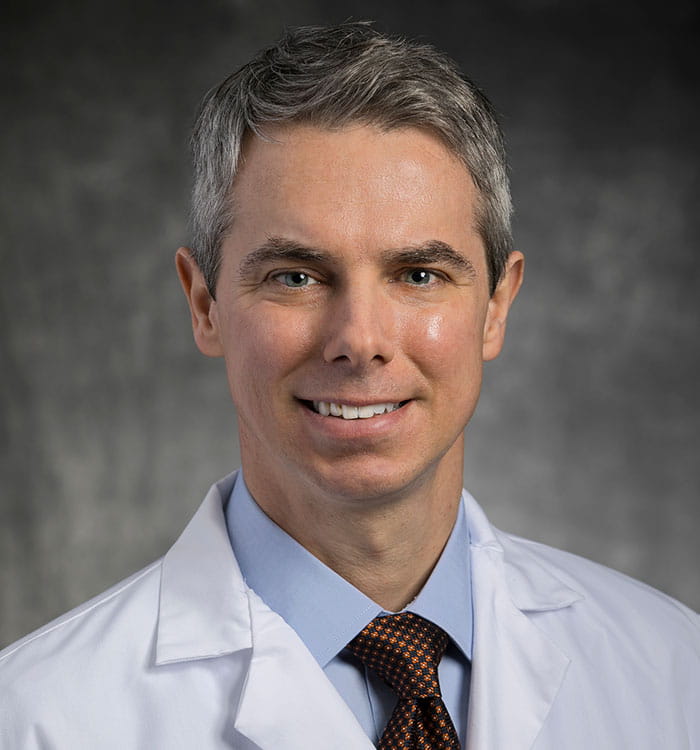
 Steven Filby, MD
“Our program growth has been greatly impacted by switching to a CTA [computed tomography angiography]-guided approach.1 By eliminating the need for TEE [transesophageal echocardiogram], we can image the appendage without general anesthesia.” In fact, the majority of University Hospitals’ LAAC patients have the procedure under conscious sedation and go home the same day.
Center of Excellence
LAAC is a minimally invasive technique to seal off the heart’s left atrial appendage, preventing blood pooling and clotting that could lead to stroke. The procedure is indicated for patients with nonvalvular atrial fibrillation (AFib) who have a suitable rationale to discontinue lifelong anticoagulant therapy.
Recognized as a Center of Excellence, the LAAC program at University Hospitals Harrington Heart & Vascular Institute is achieving higher rates of procedural success than the national average, with much lower rates of complication or device leakage. Additionally, Dr. Filby explains that patient satisfaction has driven increased demand. “We obtain imaging, plan the procedure and deliver the device during a single appointment — all within our hybrid cath lab suite,” he says. “Most patients are ready for discharge after three hours of post-procedural observation.”
Innovating Around the Coronavirus Pandemic
Conventional planning for left atrial appendage closure has relied on TEE, an invasive study in which an echo or ultrasound probe is placed down the patient’s esophagus under general anesthesia. However, early in the coronavirus pandemic, a moratorium was placed on TEE because aerosolized particulate matter could potentially transfer to healthcare providers and other patients. Experts within UH Harrington Heart & Vascular Institute collaborated on a solution that would enable them to continue LAAC in a safe manner.
“Engaging with our advanced imaging physicians, our team shifted to using CTA to ensure the morphology of the appendage,” says Dr. Filby. “CTA enables us to study the heart in a slice-like fashion in order to make sophisticated decisions about device selection and trajectory of delivery to achieve complete closure.”
Because CTA offers much more accurate images than the grainy two-dimensional grayscale of TEE, the team’s accuracy in device selection has improved from approximately 80 percent to 95 percent. “That is important because if we choose the wrong device, we have to prolong the procedure to remove and replace it,” explains Dr. Filby. “The tissue of the left atrial appendage is extremely thin, so we want to avoid any unnecessary instrumentation of this part of the heart.”
During device deployment, intracardiac echocardiography (ICE) provides high-resolution visualization. A small-caliber intravascular ultrasound probe is placed into the femoral vein at the level of the groin. “Like a small periscope, it allows us to visualize the structures inside the heart,” says Dr. Filby. “It is placed through the IV, not through the esophagus, and gives us information similar to TEE without placing anything down the patient's throat or requiring general anesthesia.”
Overcoming a Worldwide Contrast Shortage
Last spring, a worldwide shortage of iodinated contrast media halted diagnostic imaging studies at many hospitals, limiting its use to emergency diagnostic testing. “I approached my patients to say we didn’t have the necessary contrast, and patients wanted to delay their procedures,” says Dr. Filby. “Patients vote with their feet. They heard such good things from people who underwent LAAC with our CTA approach and were unwilling to have the procedure using the older, conventional way of planning.”
Once again, the team returned to the drawing board and developed a protocol using cardiac magnetic resonance imaging (MRI) as an alternative modality. UH Harrington Heart & Vascular Institute was the first center in the world to report LAAC with MRI and the first to subsequently report a series of patients. The protocol is now being used at Ohio State and other centers around the country.
Providing National Leadership
Dr. Filby and his colleagues have published the largest experience of offering same-day discharge for LAAC2 of any center nationwide. “These findings are significant because our patients have had great outcomes and satisfaction in not having to spend the night in the hospital,” he says. The team has also demonstrated improved efficiency and a 15 percent cost reduction over a conventional approach requiring overnight admission.
Leaders within UH Harrington Heart & Vascular Institute are working with physicians nationwide, providing training on their novel approaches to advanced imaging for LAAC preplanning and device deployment. “These imaging modalities provide extensive information about the structures of the heart, and the left atrial appendage in particular, and ultimately lead to a better patient experience,” says Dr. Filby. “It has been rewarding to share our workflows and strategies with our peers at other healthcare systems through lectures and publications.”
For more information or to refer a patient, contact Dr. Filby at Steven.Filby@UHhospitals.org.
1 https://pubmed.ncbi.nlm.nih.gov/36300704/
2 https://pubmed.ncbi.nlm.nih.gov/36316818/
Steven Filby, MD
Program Director, Interventional Cardiology
University Hospitals Harrington Heart & Vascular Institute
University Hospitals Cleveland Medical Center
Assistant Professor
Case Western Reserve University School of Medicine


