


Dual Energy Computed Tomography May Simplify Screening for Bronchoscopic Lung Volume Reduction
November 22, 2022
Innovations in Pulmonary & Sleep Medicine | Fall 2022
Lung volume reduction surgery (LVRS) and bronchoscopic lung volume reduction (BLVR) may help qualified patients with emphysema improve their breathing and quality of life.
LVRS involves the removal of the most damaged parts of the lung to make room for healthy lung tissue. Similarly, BLVR — one-way valves strategically placed in specific branches of the lungs — deflates damaged sections of the lungs, but without the need for surgery. Both help patients breathe more comfortably and increase their capacity for activity. University Hospitals Cleveland Medical Center is one of only a few centers in Ohio that offers both these procedures.
Patients are carefully screened to determine if they are appropriate candidates for BLVR and must undergo a thorough workup and evaluation, explains Sameer Avasarala, MD, pulmonologist at University Hospitals Cleveland Medical Center.
 Sameer Avasarala,MD
“BLVR can be an option for patients with severe emphysema whose symptoms are not well controlled with standard medical therapy,” he says. “The most important part of BLVR is picking the right target lobe(s) for the valves. We use a variety of tools to help us. Right now, during the standard workup process, we obtain a quantitative perfusion scan to get estimates of blood flow to different areas of the lung.”
New Clinical Application for Existing Technology
Dr. Avasarala received a CHEST Foundation grant to evaluate Dual Energy Computed Tomography (DECT) scans as an alternative to quantitative perfusion scans. He will be studying this with his co-investigator, Amit Gupta, MD, a DECT scanning expert in the Department of Radiology at University Hospitals Cleveland Medical Center. These DECT scans produce an iodine map that shows blood flow in the lungs.
 Amit Gupta, MD
“The main point of this project is to see how much blood flow changes in the lungs pre- and post-valve placement using dual-energy CT scans,” says Dr. Avasarala. “Every patient enrolled in the study will have a quantitative perfusion scan on file, as well. We want to explore the idea that patients may not need this scan prior to valve placement.”
“If this is the case, these patients — who are already undergoing numerous tests — can have all their screening and evaluation through a single CT scan, making it much easier for them,” says Dr. Avasarala.
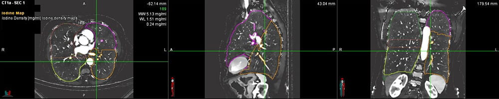 Dual Energy Computed Tomography (DECT) scan
Enrollment Open
Drs. Avasarala and Gupta will begin enrolling patients in this trial (LUng voluMe reductioN by DuAL ENergy CT Evaluation, or LUMINANCE) in November 2022. Clinicians are encouraged to refer patients for consideration.
“The ideal patient for this trial is someone with severe emphysema who is still very symptomatic with maximum medical therapy,” Dr. Avasarala says. “If the patient qualifies to receive endobronchial valves, then they qualify for this study as long as they can undergo a contrast-enhanced CT scan.”
Dr. Avasarala hopes this study continues to increase awareness of LVRS and BLVR as treatment options for patients with emphysema. If they can prove this concept, it will simplify the procedure for patients and improve the quality of comprehensive care University Hospitals delivers to patients with emphysema. Furthermore, Dr. Avasarala hopes this study demonstrates further applications for DECT scans.
Comprehensive Care for Emphysema
“When we see patients in our clinic for BLVR, we not only discuss LVRS, we also discuss lung transplantation and other treatment options,” Dr. Avasarala says. “From a clinical perspective, this study is a unique intersection between cardiothoracic imaging and interventional pulmonology and a good example of collaboration between two very distinct departments.”
For more information about this study or BLVR, call Dr. Avasarala at 216-844-3201.
Contributing Expert:
Sameer Avasarala, MD
Pulmonologist
University Hospitals Cleveland Medical Center
Assistant Professor
Case Western Reserve University School of Medicine