


Transformative Eye Research Led by UH Expands Donor Pool for Corneal Transplant Patients
June 25, 2026
UH Clinical Update | June 2026
For thousands of patients awaiting a corneal transplant, the greatest barrier to restored vision is access to donor tissue.
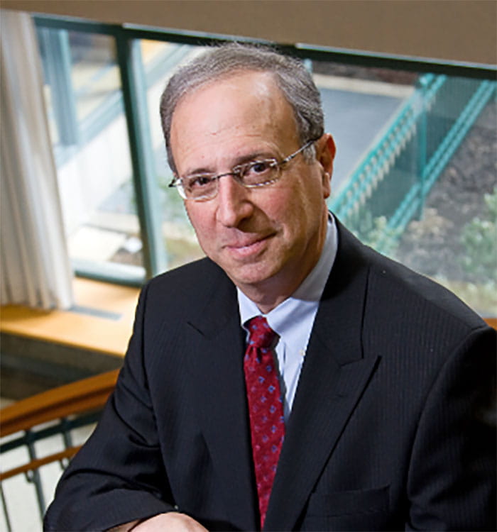 Jonathan Lass, MD
For years, many eye banks have hesitated to use corneas from donors with diabetes, concerned that the tissue might be more difficult to prepare or less likely to succeed after transplantation. That created a significantly reduced donor pool at a time when demand continues to grow with an aging population.
A groundbreaking clinical trial led by researchers at University Hospitals and Case Western Reserve University is poised to shift that paradigm.
Published in JAMA Ophthalmology in October last year, the Diabetes Endothelial Keratoplasty Study (DEKS) found that corneas from donors with diabetes perform just as well as those from non-diabetic donors, at least in the first year after surgery.
“The results were really great news,” says Jonathan Lass, MD, Charles I. Thomas Professor of Ophthalmology and Visual Sciences at Case Western Reserve School of Medicine and the study’s principal investigator. “We were expecting the more severe diabetic donors would do worse, but they didn’t.”
The concern around diabetic donors has been practical and biological. During preparation for a type of transplant known as Descemet Membrane Endothelial Keratoplasty (DMEK), surgeons must carefully peel a delicate, ultra-thin membrane, Descemet membrane, with its attached endothelial cells from the donor cornea.
“In earlier experience, tissue from diabetic donors had a higher tear rate during preparation,” Dr. Lass says. “That raised concerns that the tissue might be more fragile or less viable.”
That challenge, combined with the growing prevalence of diabetes among potential donors, is what led Dr. Lass and his colleagues to tackle what he calls “one of the next big questions that would impact eye banking and cornea transplantation.”
“We knew there was a large percentage of donors with diabetes, approaching 40% of donors in most eye banks,” he said. “What we didn’t know was to what extent we could safely use them.”
The answer required a rigorous, large-scale effort, beginning with obtaining funding.
After several attempts, the study secured funding from the National Eye Institute (NEI) of the National Institutes of Health in 2021. NIH grants are received and administered by Case Western Reserve University. The trial enrolled 1,097 patients at 28 clinical sites, involving 46 surgeons and tissue from 13 U.S. eye banks.
Participants underwent DMEK, a procedure that replaces only the innermost layer of the cornea, known as the endothelium. Two-thirds received tissue from non-diabetic donors, while one-third received tissue from donors with diabetes.
To ensure accuracy, the research team relied on more than medical records. The donor’s diabetes status was confirmed using postmortem hemoglobin A1C testing, which showed that about 10% of donors initially thought to be non-diabetic actually met the criteria for the disease.
After one year, the results were clear: graft success rates were equivalent between the two groups, 96% in the non-diabetic donor group and 97% in the diabetic donor group.
Just as important, the severity of diabetes in the donors — from mild cases managed with oral medication to advanced disease with complications including retinopathy —had no effect on outcomes. “We found no difference at all,” Dr. Lass says. “Diabetes doesn’t make a difference, nor does the severity.”
The study was lauded for being well-designed, well-executed and highly impactful, which led to JAMA’s recognition of the clinical trial, as the Clinical Trial of the Year in 2025 for ophthalmology. Co-investigators in the study included Loretta Szczotka-Flynn, OD, PhD (Ophthalmology); Beth Ann Benetz, MA (Ophthalmology); Baha Arafah, MD, (Medicine); Vincent Monnier, MD (Pathology) and Sudha Iyengar, PhD, (PQHS).
Expanding the use of this tissue could help alleviate a persistent global shortage.
The study did identify one important distinction: tissue from diabetic donors is slightly more prone to tearing during preparation, by about 3-4%.
But once successfully prepared, outcomes are equivalent.
“The message is, yes, there is a slightly higher risk during preparation,” Dr. Lass said. “But if you can successfully peel it, the cells do well.”
As a result, many eye banks are rethinking their use of donors with diabetes, expanding their pool of donors for the corneal transplant procedures they are supporting.
The work is continuing. With continued NEI funding, which is part of a $17 million total award, the team will follow patients for five years to determine whether the results hold over time.
Future research will also explore genetic factors, including polygenic risk scores that could help predict graft outcomes based on donor and recipient genetic characteristics, an approach that could be a first in managing conditions like Fuchs dystrophy.
For Dr. Lass, who has been with UH since 1979, the DEKS trial brings together his decades of clinical and research experience.
As a corneal transplant surgeon and a longtime leader at University Hospitals, he has spent his career advancing the science of eye banking, from preservation techniques to transplant outcomes.
“This trial brings together all of my interests,” he said. “As a surgeon, you impact one patient at a time. But with clinical research like this, the impact is much broader.”
The journey from concept to publication took more than a decade, and he describes it as both challenging and deeply rewarding.
“You have to have tremendous perseverance,” he said. “But in the end, knowing this could help restore sight to so many more patients, that’s what makes it worthwhile.”
A companion study, conducted by the Cornea Image Analysis Reading Center at Case Western Reserve and University Hospitals, examined what happens to endothelial cells after transplant.
These cells play a crucial role: they pump fluid out of the cornea to keep it clear. Unlike many other cells in the body, they do not regenerate.
Using advanced imaging techniques, researchers tracked changes in these cells over time. At one year, endothelial cell loss was identical — 28% — in both groups.
“These results support no restrictions on the use of tissue from donors with diabetes for DMEK,” Dr. Lass said. “That has the potential to expand the donor pool significantly.”