


Custom 3-D Printed Airway Stents Markedly Improve Pulmonary Patient Outcomes
October 25, 2023
University Hospitals Cleveland Medical Center early adopter of this approach
Innovations in Pulmonology, Critical Care & Sleep Medicine | Fall 2023
Commercial stents have long been the gold standard for treating patients with benign airway stenosis, says Benjamin Young, MD, Director, Interventional Bronchoscopy, University Hospitals Cleveland Medical Center. “We use stents to treat patients with nonmalignant airway disease who are not candidates for surgery,” he says. “These patients have diseases that lead to airway instability or narrowing. This can result in the inability to breathe effectively, or to clear secretions, which can set up patients for complications such as pneumonia.”
 Benjamin Young, MD
Commercial stents have long been the gold standard for treating patients with benign airway stenosis, says Benjamin Young, MD, Director, Interventional Bronchoscopy, University Hospitals Cleveland Medical Center. “We use stents to treat patients with nonmalignant airway disease who are not candidates for surgery,” he says. “These patients have diseases that lead to airway instability or narrowing. This can result in the inability to breathe effectively, or to clear secretions, which can set up patients for complications such as pneumonia.”
Airway stents are easy to place and remove through bronchoscopy. While immediate results are generally positive after stent placement, over time, patients frequently develop complications that can diminish the efficacy, including inflammation, mucus formation and mechanical rubbing, which causes tissue formation and may occlude the lumen of the stent.
3-D Stents Offer New Therapeutic Options
“The biggest contributor to the development of these complications is that the stents don’t fit all that well,” Dr. Young says. “Commercially available stents are either straight cylinders or in the shape of a Y, with straight limbs. There is no accounting for the airway curvature, branching or changing diameter as the airway extends deeper into the lung. Poor fit leads to an exacerbation of problems.”
In late 2019, the U.S. Food and Drug Administration approved custom 3-D patient-specific airway stents. The team at UH Cleveland Medical Center placed the first of these in November 2020 and has since utilized this technology for many other patients with notable outcomes.
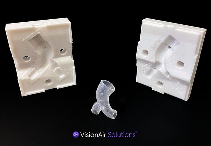
The process of creating a custom stent begins with a high-resolution CT scan. The image is uploaded, and a computer software program creates a 3-D reconstruction of the airway. Using the software, the physician then designs a stent that matches the specific unique parameters of the patient’s airway, accounting for airway diameter and length, branching, wall thickness and angulation.
From this virtual prescription, a custom mold is 3-D printed and medical-grade silicone is injected to create a stent that exactly fits the patient’s airway. The stent is implanted under sedation in an outpatient setting by a thoracic surgeon or interventional pulmonologist, and patients generally go home the same day.
UH Cleveland Medical Center is Early Leader in 3-D Airway Stenting
In 2018, Dr. Young and colleagues published the results of the first-in-human clinical experience with two patients with Granulomatosis with polyangiitis (formerly called Wegener’s granulomatosis) who had not responded to standard therapies. They compared patients’ airways six months prior to 3-D stent implantation to a year after implantation and found patients had improved durability and improved patient-reported symptoms, which led to a reduced need for stent changes and modifications. “UH was one of the early adopters of this technology,” Dr. Young says. “Now that we are using 3-D stents in clinical practice, we find patients are responding favorably. The stents are well tolerated, and patients show a marked improvement in breathing and quality of life.”
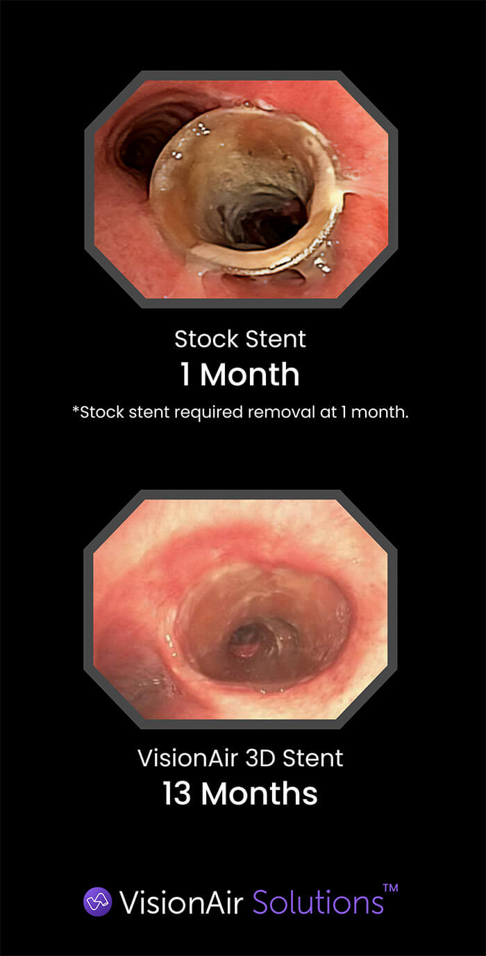
The silicon used to make the stents does degrade over time as they are exposed to air and mucus in the lungs and patients’ airways can also change over time. The stents should be replaced about every 12 months.
“Patients who have airway disease requiring a stent often have inflammatory disease and stenosis,” says Dr. Young. “We’re finding that with these better-fitting stents, there’s decreased inflammation, so the airways change for the better. Furthermore, in patients who have tracheobronchomalacia, the airways tend to dilate over time, so patients may need a larger stent.
“3-D stents are really a game changer in airway stenting and we’re trying to make more clinicians aware of this option,” Dr. Young says. “It’s a good tool for treating airway obstruction and offers clinicians more opportunities to treat patients who don’t respond to standard therapy or who experience complications with commercial airway stents.”
For more information about 3-D airway stents, or to refer a patient for consultation, contact Dr. Young at 216-541-1521 or Benjamin.Young@UHhospitals.org.
Contributing Expert:
Benjamin Young, MD
Director, Interventional Bronchoscopy
University Hospitals Cleveland Medical Center
Associate Professor
Case Western Reserve University School of Medicine