


First-in-Country Magnetic Resonance Fingerprinting for Opioid-Exposed Infants at UH Rainbow
October 25, 2021
![]()
UH Rainbow only site in the U.S. using next-generation imaging in study of infants with Neonatal Abstinence Syndrome
Innovations in Pediatrics | Fall 2021
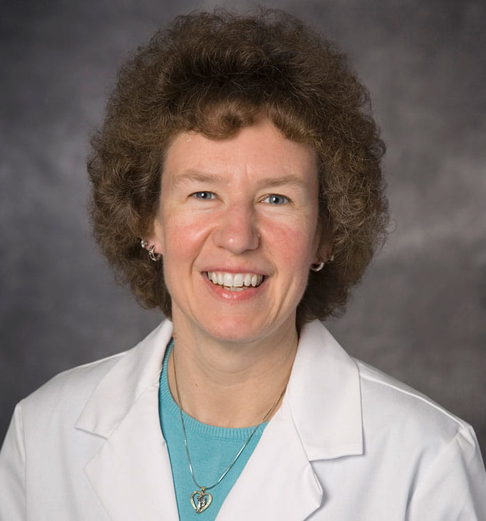 Deanne Wilson-Costello, MD
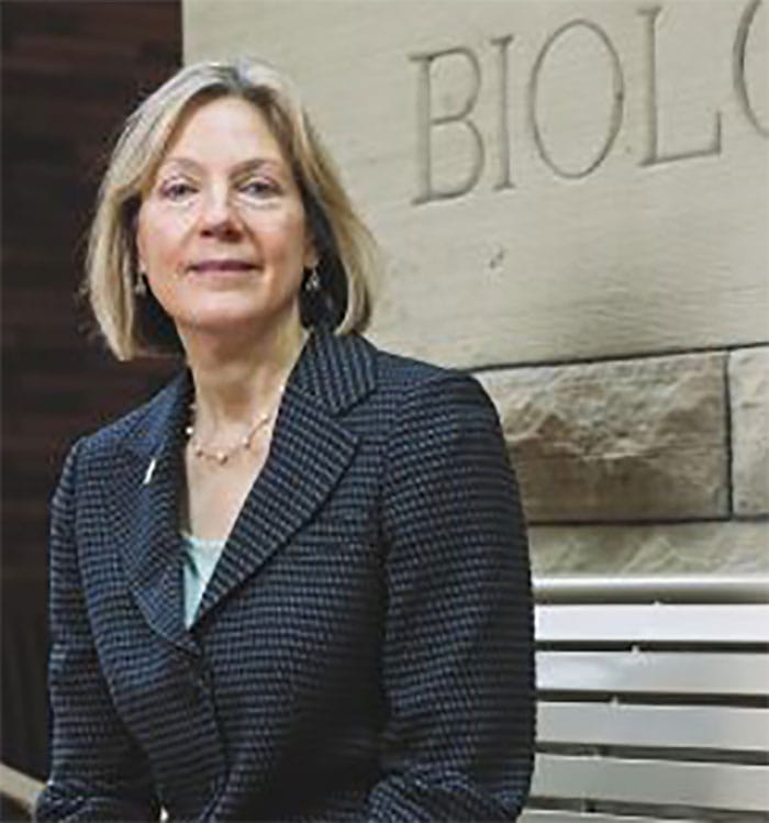 Lynn T. Singer, PhD
Magnetic resonance fingerprinting (MRF), first described in the journal Nature in 2013 by engineers at Case Western Reserve University School of Medicine and University Hospitals, represents a revolution in imaging. With this new technology, radiologists here are able to generate immediate and simultaneous values for T1 and T2 relaxation times for each pixel in the image, layering quantitative values onto the qualitative information available from typical MR. These tissue characteristics describe the environment of each particular proton, such as fat or water. The signal from each pixel is matched against a “dictionary” of all possible signals, and the match yields the underlying quantitative values that went into creating the dictionary entry, even when the signal itself is “noisy.” It’s akin to distinguishing the letter “A” from the letter “B” on a page, even when parts of the letter are faint or missing, or to being able to correctly identify “The Star-Spangled Banner” on a radio with a lot of static, just based on the words “say” and “see” and a fragment of melody.
To date, the main advantage of MRF in practice has been its ability to distinguish between tissue types in adult patients, such as primary from secondary brain tumors and the more aggressive glioblastoma multiforme from oligodendrogliomas. But now, a new population of patients at UH Rainbow Babies & Children’s Hospital is benefiting from MRF and one of its key advantages – its speed.
UH Rainbow neonatologist Deanne Wilson-Costello, MD, and Lynn T. Singer, PhD, Professor of Population & Quantitative Health Sciences at Case Western Reserve University School of Medicine, are employing MRF in a new study of infants with Neonatal Abstinence Syndrome (NAS), looking to better understand structural changes in the infant brain that may accompany the condition.
For Dr. Wilson-Costello, the best part of the new study is that MRF can yield so much information in so little time.
“Because the NAS babies are often times so jittery and difficult to console, and hyper-responsive to stimulation, the MRI is a nightmare for them,” she says. “It is loud and creates a lot of sensory overload. The beauty of MRF, instead of taking one image and then varying a parameter, it varies multiple parameters in the MRI at the same time, so it acquires an image very rapidly, between a minute and 10 minutes, as opposed to 35 to 40 minutes. The more time we can save in the scanner, the less consoling we have to do for an agitated NAS baby. So here it’s even more valuable than in other applications.”
“Since a normal scan takes up to an hour, scheduling, completing scans and obtaining cooperation from families to do so for research purposes is quite difficult,” adds Dr. Singer. “MRF significantly reduces the amount of time required and can revolutionize our ability to obtain this much needed information about brain development, leading to more successful scans and retention of subjects.”
Dan Ma, PhD, Assistant Professor of Biomedical Engineering at Case Western Reserve University and one of the experts behind the development of MRF, outlines other advantages.
“MRF provides reproducible and quantitative maps of key tissue properties,” she says. “These quantitative tissue property maps can be used to analyze or track developmental changes of infant and baby brains longitudinally. For example, studies have identified that quantitative T1, T2 and myelin water fraction values are rapidly changing during the first few years of life, which are closely linked to neurodevelopment, cognitive abilities and nutrition level during the brain growth, while deviation of the normal trajectory might indicate alternations of brain development. Typical MR scans are qualitative measurements, which do not provide such quantitative maps for longitudinal studies.”
The new NAS/MRF study at UH Rainbow is an ancillary study to the federally funded ACT NOW OBOE study (Advancing Clinical Trials in Neonatal Opioid Withdrawal Syndrome: Outcomes of Babies with Opioid Exposure), supported by the National Institutes of Health at UH Rainbow and three other sites nationwide. However, UH Rainbow is the only site to offer MRF. UH neuroradiologist Chaitra Badve, MD, provides expert support in conducting the MRF scans.
Dr. Wilson-Costello says that because their study is an ancillary to the larger ACT NOW OBOE, she and Dr. Singer are able to make instructive comparisons between different groups of patients.
“When we got the grant for the OBOE study that looks at opiate exposures, we had a cadre of kids who were already set to get standard MRIs, so it gave us the ability to put in the fingerprinting and be able to compare the pictures of a standard MRI to the information we get with MRF,” she says. “The beauty of the OBOE cohort is that there are control infants as well as exposed infants. So you can actually look at what the fingerprinting is going to look like in an opioid-exposed kid and a control kid.”
The goal with all of this research, Dr. Wilson-Costello says, is laying the groundwork for developing different approaches to treating NAS infants more effectively.
“When we have a better understanding of the brain structure, we’ll be better able to target interventions for areas that may be damaged and may be better able to prevent the damage from happening,” she says. “If we have antenatal interventions aimed at a specific area, we could potentially improve outcomes.”
However, she says she expects the benefits of MRF technology will not just be limited to NAS infants, but rather ultimately to all pediatric patients who need an MR scan.
“It’s really going to have great application to all of pediatrics if it flies,” she says. “It’s collaborative from basic research to clinical application to a helpful tool for potentially all centers.”
For more information or to contact UH Rainbow, email Peds.Innovations@UHhospitals.org.
Contributing Experts:
Deanne Wilson-Costello, MD
Neonatologist, Division of Neonatology
Director, High Risk Follow-up Program
UH Rainbow Babies & Children's Hospital
Professor of Pediatrics
Case Western Reserve University School of Medicine
Lynn T. Singer, PhD
Professor of Population & Quantitative Health Sciences
Case Western Reserve University School of Medicine
Dan Ma, PhD
Assistant Professor of Biomedical Engineering
Case Western Reserve University