


OCT Bringing Heart Imaging to Light
January 26, 2017
Harrington Heart & Vascular Institute Innovations – Winter 2017 – View Full PDF
 Hiram Bezerra, MD, PhD
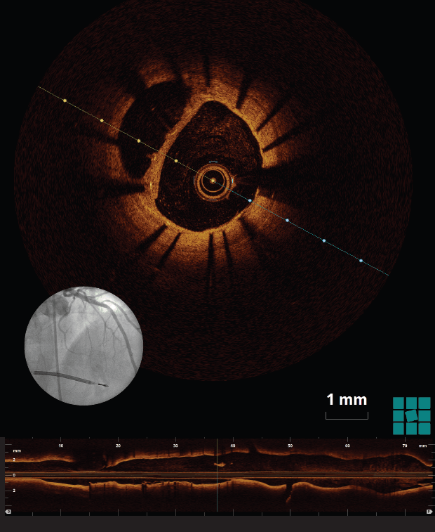
Optical coherence tomography (OCT), a catheter-based invasive imaging system, uses light instead of ultrasound to produce high-resolution, real-time images of coronary arteries and deployed stents for patients with cardiovascular disease. OCT allows cardiologists to see details inside blood vessels that were never seen before, and the resulting images are almost to the level of a microscope in terms of resolution. The visualization can be more than 10 times the detail of intravascular ultrasound.
A team at University Hospitals Harrington Heart & Vascular Institute using intravascular OCT was able to rule out intraluminal thrombus, and thus further intervention, following a stent placement in the left anterior descending artery. Their findings, “Optical coherence tomography assessment ruled out the need for intervention in a ‘hazy’ angiographic image,” were published in The International Journal of Cardiovascular Imaging in September 2016. The authors are Mohamad Soud, MD; Guilherme F. Attizzani, MD; Daisuke Nakamura, MD; Gabriel Tensol Rodrigues Pereira, Research Fellow; Setsu Nishino, MD, PhD; and Hiram G. Bezerra, MD, PhD. All are affiliated with UH.
They found that OCT’s high resolution proved more accurate than intravascular ultrasound (IVU) in identifying intraluminal thrombus and that, compared to coronary angioscopy, OCT has 100 percent sensitivity versus 33 percent for IVUs in detecting intracoronary thrombus.
This advanced technology is shaping the future of cardiovascular imaging, and the Cardiovascular Imaging Core Laboratory at UH is leading the way in its use.
“For several years, OCT has been a routine part of our arsenal for coronary prevention,” says Hiram Bezerra, MD, PhD, the study’s senior author, Director, Cardiac Catheterization Laboratories, University Hospitals Cleveland Medical Center; Associate Professor of Medicine, Case Western Reserve University School of Medicine. “We use intravascular imaging in approximately 80 percent of our cases.” Nationally OCT is used in roughly 5 to 10 percent of all coronary interventions.
OCT is useful as a diagnostic tool, offering excellent tissue characterization for plaque components and planning interventions. Therapeutically, this modality facilitates interventions by offering the most precise dimensions for device selection, which optimizes results after stenting by revealing things such as stent expansion and positioning. Biodegradable stents need even more precision in implantation than other types of stents do, and OCT offers that precision. OCT allows doctors to accurately measure luminal architecture and gain insights regarding stent placement and, in the case of biodegradable stents, information about the time it takes for them to dissolve.
UH has been a pioneer in research and clinical use of OCT since its investigational phase. A UH team published a comprehensive review of intracoronary OCT in the Journal of the American College of Cardiology: Cardiovascular Interventions in 2009. The paper described the technical aspects of OCT and highlighted its then-emerging research and clinical potential. The authors were Dr. Bezerra; Marco A. Costa, MD, PhD; Giulio Guagliumi, MD; Andrew M. Rollins, PhD; and Daniel I. Simon, MD. UH Cleveland Medical Center was the first U.S. site to teach doctors to perform OCT and continues to be a training site.
For more information or to refer a patient, call 216-844-3800 or email HVInnovations@UHhospitals.org.