Sound Solution
January 01, 2016
Auditory brainstem implant restores hearing to normal range for teenager born without cochleas
Innovations in Otolarngology - Head and Neck Surgery Winter 2016 Download PDF
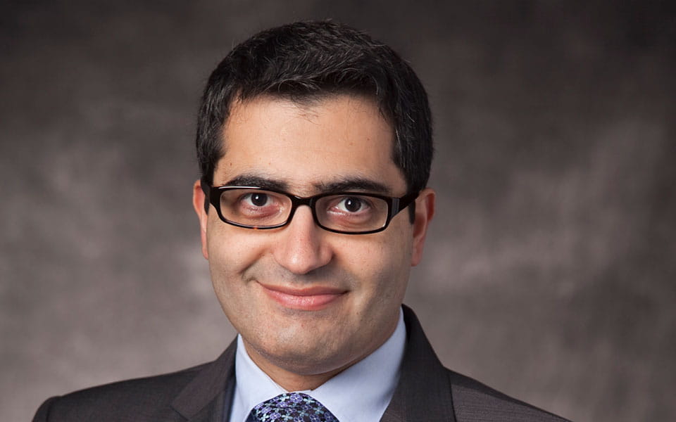
 Maroun Semaan, MD
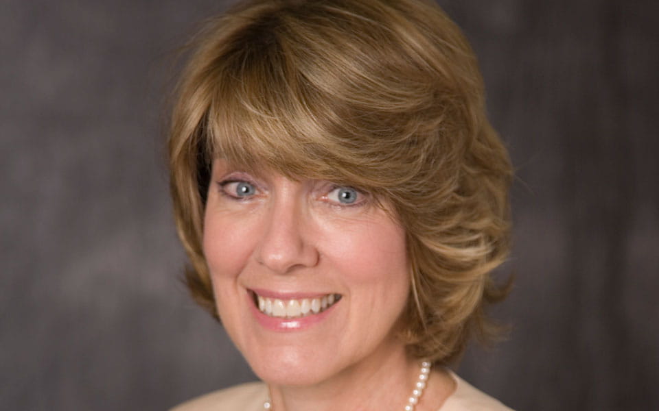
 Gail Murray, PhD, CCC-A
Auditory brainstem implantation (ABI) surgery has typically been reserved for adult patients with bilateral vestibular schwannomas caused by neurofibromatosis type 2 (NF-2), as a means of restoring hearing. However, over the past 10 years, indications for the procedure have evolved. Although pediatric ABI surgery is still being studied in clinical trials, specialists are increasingly offering the procedure to pediatric patients and their families.
“ABI surgery is now indicated for people who were born deaf or with profound hearing loss, either because they never had cochleas develop or have only small, rudimentary remnants of cochleas that cannot receive a cochlear implant,” says Maroun Semaan, MD, Associate Director of Otology and Neurotology and Director of the Cochlear Implant Program, University Hospitals Case Medical Center and UH Ear, Nose & Throat Institute, and Assistant Professor of Otolaryngology, Case Western Reserve University School of Medicine. “ABI surgery is now also indicated for those who were born without an auditory nerve (cochlear nerve aplasia) or a very thin auditory nerve (cochlear nerve hypoplasia).”
A multidisciplinary team at UH recently had the opportunity to put these expanded indications into practice. Dr. Semaan, UH Ear, Nose & Throat Institute Director Cliff Megerian, MD, neurosurgeon Nicholas Bambakidis, MD, and audiologist Gail Murray, PhD, CCC-A, provided an auditory brainstem implant to a 14-year-old girl born without cochleas – the first pediatric patient in Ohio to undergo such a procedure.
The patient had developed language using the total communication approach of voicing and American Sign Language and had used a vibrotactile device for picking up on environmental cues. “She had a really nice foundation when it came to language,” Dr. Murray says. “It made it much easier to work with her and explain concepts for her to understand and follow.”
Before the surgery, Dr. Murray and her audiology team worked with the patient and her family to set reasonable expectations. “We wanted them to understand that this was going to be hard and tedious and would require many visits,” she says. “They all needed to be on board with it. Everyone had to understand what was to come.”
In performing the surgery, Dr. Bambakidis and Dr. Semaan opted for the retrosigmoid approach to reach the brainstem, as opposed to the more traditional translabyrinthine craniotomy. The most challenging test came in placing the internal electrode pad in the correct location on the cochlear nucleus.
“With the ABI, it’s a bit of blind/nonblind technique, where you apply the electrodes on the dorsal surface of the fourth ventricle – the approximate location of the cochlear nucleus,” Dr. Semaan says. “You don’t know exactly where it is, how well developed it is and what to expect. We didn’t really know what was going to happen when we put that implant in the brainstem. Was the cochlear nucleus still there? Did it develop? Did it degenerate? Was there some neuronal plasticity involved with it being used for nonauditory perception or function?
Fortunately, intraoperative electrical auditory brainstem response (EABR) testing confirmed that the implant was indeed in the right place. “That’s very important,” Dr. Semaan says. “Otherwise we can stimulate nonauditory centers of the brainstem, which can cause side effects that are quite unpleasant and can sometimes be dangerous.”
“When we activated it, we were all happy to see that she had auditory perception without a doubt,” Dr. Semaan adds. “We cheered for her, but we were also very interested from a neurophysiological perspective. Even when you had the end organ not developing, the ‘docking structure,’ in this case, the cochlear nucleus, did not go away, disappear or ramp down. It was there waiting for one day to receive the signal.”
After the internal electrode pad was placed and verified, Dr. Murray and her team tested the ABI on all 20 of its electrode channels. “We kind of bracketed them based on their location on the brainstem,” she says. “When we got response on one, we jumped to another area. We did that regional testing and filled in the gaps afterward.”
The team activated the device about three months post-surgery. The first sound the patient heard was her father’s voice (see sidebar). Intensive sessions to map the patient’s responses to auditory stimuli soon followed.
“The early stages of mapping were for us to see where she had a response, hopefully an auditory one, and then making it comfortably loud,” Dr. Murray says. This testing revealed that four of the 20 channels should be turned off. Some were creating unwanted nonauditory responses, and others were unpleasant to the patient in how they sounded.
Later mapping focused on having the patient rank pitches according to their frequency. “Pitch ranking is the most difficult task,” Dr. Murray says. “The auditory system is organized in a tonotopic manner, from the inner ear, through the lower brainstem to the cortex of the brain. The goal is to assign the electrodes on the ABI pad the frequency that is perceived and organize the electrodes in a way that best matches the tonotopic organization of the patient’s brainstem and auditory pathway.
“Take the word ‘scissors,’ which is a high-pitched word,” Dr. Murray adds. “If I don’t have the electrodes that are assigned those high frequencies in the area where she perceives it as high frequency, she’ll perceive it as something completely different. If we have this sequenced incorrectly, it will impede her ability to start learning the meaning of words.”
After many hours of mapping sessions and acclimating to sounds, the patient is now hearing in the normal range. Dr. Murray attributes some of this success to new external speech processor technology. The updated processor is lighter and thinner than its predecessor. Plus, it automatically adjusts the digital sound algorithm based on the needs of the sound environment, without the patient having to manually change programs.
“For me, that’s one of the most exciting things,” Dr. Murray says. “It’s what brought her from mild hearing loss level into normal hearing level. After the first adjustments, I’m sure she heard her dog bark and her father’s deep voice, but a lot of different voices, she would not have been able to appreciate unless they were raised and shouting. But now that we’re finished mapping, there is a big difference, which is exciting to see. Now her responses are in normal hearing range.”
The patient is continuing with auditory verbal therapy to develop listening and speaking skills.
For both Dr. Semaan and Dr. Murray, outcomes like these point to the importance of a multidisciplinary approach to complex issues.
“This work only happens because of the close collaboration of many departments,” Dr. Semaan says. “There was a whole team of experts involved, and everybody had to do their best work.”
For more information on this case, email Maroun.Semaan@UHhospitals.org or Gail.Murray@UHhospitals.org.
Patient’s Story Reaches Millions
More than 4 million people worldwide have witnessed the moment when the young ABI patient at University Hospitals heard her father’s voice for the first time. Digital outlets that published her story include USA Today, Huffington Post, People.com, Daily Mail and CNN. The original video, hosted on University Hospitals YouTube page, generated approximately 900,000 impressions on YouTube alone.
Tags:


