


Cornea Image Analysis Reading Center (CIARC)
Expert Evaluation of Corneal Imaging for Clinical Trials
The Cornea Image Analysis Reading Center provides specialized grading and analysis for studies involving external, corneal and anterior-segment imaging.
Contact Us Today
Call: 216-844-3615
Email: EIARC@UHhospitals.org
or fill out our online form.
The Cornea Image Analysis Reading Center services include evaluation of epithelial healing, endothelial safety and toxicity, drug and device performance, and surgical efficacy. Additionally, the Cornea Image Analysis Reading Center conducts ocular safety studies for non-ophthalmic clinical trials, ensuring that systemic drug and device studies include rigorous evaluation of potential ocular effects.
Our dual-grading system, involving two independent graders with a third experienced grader adjudicator, as needed, ensures objectivity and accuracy. The CIARC’s web-based image management platform allows seamless, multi-modal submission, transfer, and tracking of all study images and data.
Areas of Expertise
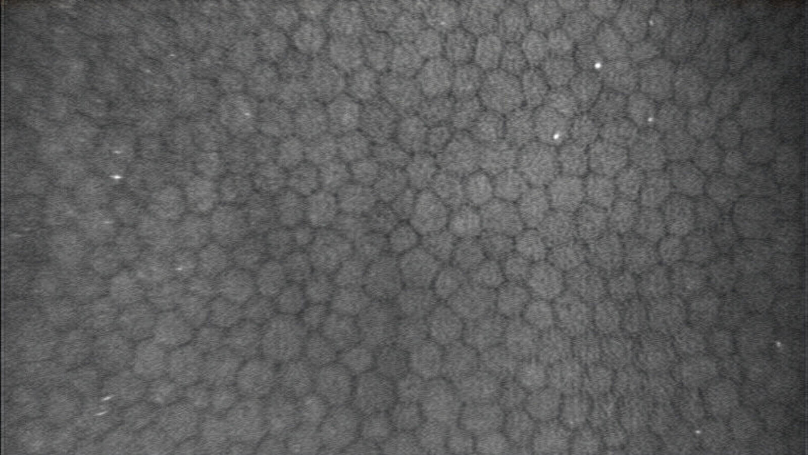
- Corneal endothelial cell density and morphology
- Guttae analysis
- Corneal epithelial staining and defect measurement
- Confocal image analysis
- Safety assessments for drugs, devices and surgical procedures
- IOL positioning assessment
- Non-ophthalmic drug safety ocular studies
At the Forefront of New Technologies
In partnership with Case Western Reserve University’s Department of Biomedical Engineering, CIARC is advancing machine learning applications that move beyond traditional imaging software to transform lid, cornea and anterior segment image analysis. Current projects focus on developing AI-driven algorithms for the automated assessment of endothelial cell density and morphology of the donor cornea endothelium, as well as guttae in Fuchs endothelial corneal dystrophy. The CIARC in conjunction with Case Western Reserve University’s Department of Biomedical Engineering and the Eversight Eye Bank recently was awarded a High Impact grant from the Eye Bank Association of America to advance this work for the donor corneal endothelium.

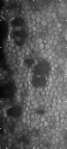
In addition, CIARC researchers are using AI-based analysis of specular microscopy images to more precisely and objectively track the progression of Fuchs endothelial corneal dystrophy, with particular focus on the quantification of guttae and endothelial cell morphology.
Finally, with advances in refractive IOLs and their long term performance, the CIARC has developed image analysis procedures to longitudinally monitor IOL position stability (tilt, rotation).
These efforts represent a critical step toward integrating AI and image-based biomarkers into clinical and translational corneal research enhancing predictive capabilities, accelerating study workflows, and refining standards for objective image interpretation.