


Eye Image Analysis Reading Centers at University Hospitals
Our Commitment
The University Hospitals Eye Image Analysis Reading Centers are dedicated to advancing ophthalmic science through rigorous data integrity, innovative technologies, and collaborative partnerships. Equipped to receive and analyze multiple imaging modalities, our centers deliver reliable, high-quality results that support regulatory success and improve patient outcomes worldwide.
Contact Us Today
Call: 216-844-3615
Email: EIARC@UHhospitals.org
or fill out our online form.
Why Work with Us
The Eye Image Analysis Reading Centers provides comprehensive image analysis and clinical trial support for studies examining the effects of ophthalmic diseases and therapies on the anterior segment and posterior segment.
30+ years of multi-site clinical trial experience
Tailored imaging protocols and data acquisition systems
Seamless receipt and review of every eye image modality
Rapid, reproducible analysis and reporting
Direct medical director oversight for data integrity
Fully compliant with GCP, ICH, FDA, HIPAA, and 21 CFR Part 11 standards
Founded in 1989, the Eye Image Analysis Reading Centers at University Hospitals Eye Institute and Case Western Reserve University unite two specialized centers—the Cornea Image Analysis Reading Center (CIARC) and the Retina Diseases Image Analysis Reading Center (REDIARC). Together, they deliver expert, standardized image grading and analysis support to assess the effects of drugs, devices, diseases and procedures on both the anterior and posterior segment.
For more than three decades, the Cornea Image Analysis Reading Center has collaborated with national and international investigators in National Institutes of Health-funded federal, and industry-sponsored clinical trials, helping to define standards for anterior segment imaging. Our work informs innovations in diagnostics, device safety, and therapeutic development across the field of ophthalmology. The Retina Diseases Image Analysis Reading Center similarly has supported industry-sponsored clinical trials that have made major advances in retina disease therapeutics.
Watch Now: Learn More About Our Services

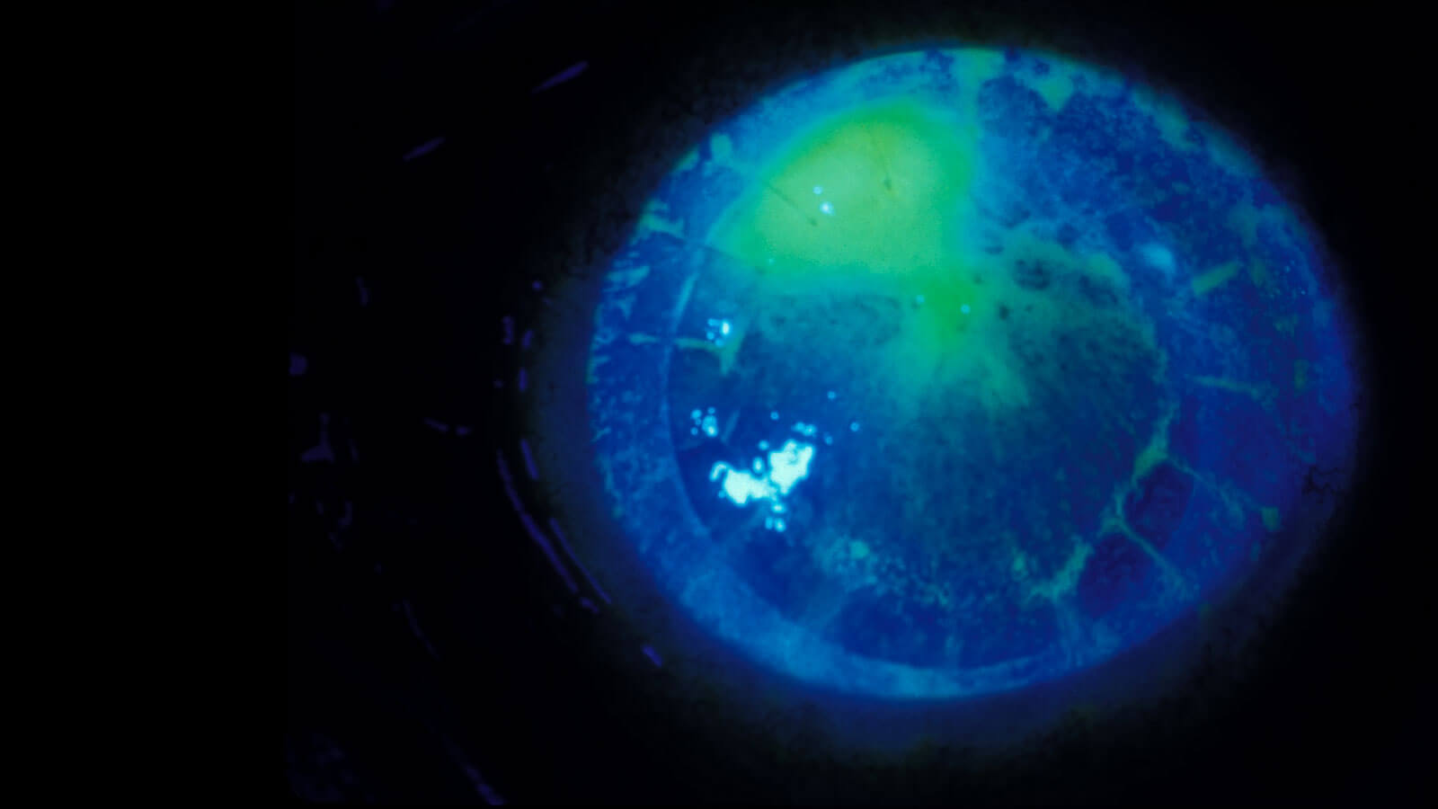
Cornea Image Analysis Reading Center (CIARC)
The CIARC provides expert image analysis and clinical trial support for studies involving the cornea and anterior segment. With decades of experience, the CIARC delivers standardized, dual-graded data and partners with academic and industry leaders to advance corneal and anterior segment research and imaging innovation.
Retina Diseases Image Analysis Reading Center (REDIARC)
The Retina Diseases Image Analysis Reading Center specializes in evaluating retinal pathology using fundus photography, fluorescein angiography, autofluorescence and OCT. This center supports national and international clinical trials assessing therapies for diabetic retinopathy, macular degeneration and uveitis, ensuring precise and reproducible imaging outcomes.

Full Range of Ophthalmic Image Analysis and Research Support
The Eye Image Analysis Reading Centers offer a complete range of ophthalmic image analysis and clinical research support services for a variety of ophthalmic conditions of the anterior and posterior segment and delivers precise, reproducible data that drives discovery and regulatory success.
Our expert readers assess parameters such as epithelial integrity including staining and defects, endothelial cell morphology, angle configuration, IOL positioning, lid abnormalities, and alterations in retinal structure in disease using modalities including external imaging, confocal microscopy, fundus photography, fluorescein angiography, meibography, optical coherence tomography, and specular microscopy. These standardized, protocol-driven assessments ensure consistency and accuracy across multicenter studies and imaging systems. They are supported by novel AI-driven image analysis methods under development.
Core Capabilities
- Study protocol design and consultation
- Imaging modality optimization and certification
- Site and technician training (on-site and remote)
- Data acquisition, grading and statistical analysis
- Quality control and adjudication
- Regulatory documentation and manuscript support
Technical Services
- Secure web-based image submission and storage
- Multi-modal portal capabilities for receipt, storage and sharing with sponsor
- Rapid image validation and feedback
- Device calibration and standardization across systems
- Quantitative and qualitative image interpretation

Partnering to Advance Ophthalmic Science Through AI and Innovation
The Eye Image Analysis Reading Centers are redefining ophthalmic research through advanced imaging technologies and artificial intelligence. In partnership with Case Western Reserve University, our team has been developing AI-driven tools that automate image analysis, detect early disease changes, and predict outcomes measured against standard image analysis methods. Combining decades of clinical expertise with cutting-edge analytics, we deliver timely, accurate, and reproducible data empowering study teams to accelerate the discovery and validation of next-generation ophthalmic therapies.
Key Metrics
Over
Over
Through long-standing partnerships with federal agencies, CROs, and industry sponsors, we help design and execute clinical trials that meet regulatory standards, generate robust, reproducible data and accelerate innovation in drug, device and surgical development.
All work is governed by the University Hospitals Institutional Review Board, accredited by the Association for the Accreditation of Human Research Protection Programs (AAHRPP).