Chorionic Villus Sampling (CVS)
What is chorionic villus sampling?
Chorionic villus sampling (CVS) is a prenatal test. It is done by taking a sample of tissue from the placenta. The sample is tested for chromosomal abnormalities and some other genetic problems. The placenta is an organ in the uterus that send blood and nutrients from the mother to the baby.
The chorionic villi are tiny fingers of placental tissue. They have the same genetic material as the baby. Testing may be done for other genetic problems. This depends on the family history and the types of lab testing that are available.
CVS is usually done between the 10th and 12th weeks of pregnancy. CVS does not give information on neural tube defects or myelomeningocele (also known as spina bifida). For this reason, women who have CVS also need a follow-up blood test between 16 to 18 weeks of their pregnancy. This test is to screen for neural tube defects.
There are 2 types of CVS procedures:
-
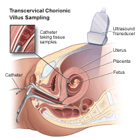
Transcervical. A catheter is put through the cervix into the placenta to get the tissue sample.
-
Transabdominal. A needle is put through the belly and uterus into the placenta to get the tissue sample.
Another test that may be used to diagnose genetic and chromosomal defects is amniocentesis. This test does give information on neural tube defects.
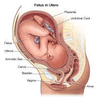
Anatomy of the baby in the uterus
-
Amniotic sac. This is a thin-walled sac that surrounds the baby during pregnancy. The sac is filled with amniotic fluid. This is liquid made by the fetus. The sac also contains the amnion. This is the membrane that covers the fetal side of the placenta. This protects the baby from injury and helps to control the temperature of the baby.
-
Anus. This is the opening at the end of the anal canal.
-
Cervix. This is the lower part of the uterus that dips down into the vagina. It is made of mostly fibrous tissue and muscle. It has a circular shape.
-
Fetus. This is a term used to describe an unborn baby from the 8th week after fertilization until birth.
-
Placenta. This is a flat organ inside the uterus that only grows during pregnancy. It sends nutrients and other substances between the baby and mother. The placenta allows the baby to take in oxygen, food, and other substances. And it lets the baby get rid of carbon dioxide and other wastes.
-
Umbilical cord. This is a rope-like cord that connects the baby to the placenta. The umbilical cord has 2 arteries and a vein. They carry oxygen and nutrients to the baby and waste products away from the baby.
-
Uterine wall. This is the wall of the uterus.
-
Uterus (also called the womb). This is a hollow, pear-shaped organ in a woman's lower belly. It sits between the bladder and the rectum. It sheds its lining each month during menstruation. When a fertilized egg (ovum) becomes implanted in the lining, a baby grows.
-
Vagina. This is a canal behind the bladder and in front of the rectum. It forms a path from the uterus to the vulva.
Reasons for the procedure
Chorionic villus sampling may be used for genetic and chromosome testing in the first trimester of pregnancy. Reasons that a woman might elect to have CVS include:
-
A previous child with, or family history of, a genetic disease, chromosomal abnormalities, or metabolic disorder
-
Maternal age over 35 years by the pregnancy due date
-
Risk of a sex-linked genetic disease
-
Previous ultrasound with abnormal results
-
Abnormal cell-free DNA test
There may be other reasons for your healthcare provider to advise a chorionic villus sampling.
Risks of the procedure
All procedures have some risks. Some risks of this procedure include:
-
Cramping, bleeding, or leaking of amniotic fluid (water breaking)
-
Infection
-
Miscarriage
-
Preterm labor
-
Limb defects in infants, a higher risk in CVS done before 9 weeks (rare)
People who are allergic to or sensitive to medicines or latex should tell their healthcare provider.
Women with twins or other multiples will need sampling from each placenta in order to study each baby.
There may be other risks depending on your overall health. Talk about any concerns with your healthcare provider before the procedure.
Certain factors or conditions may interfere with CVS. These factors include, but are not limited to:
-
Pregnancy earlier than 7 weeks or later than 13 weeks
-
Position of the baby, placenta, amount of amniotic fluid, or mother's anatomy
-
Vaginal or cervical infection
-
Samples that are inadequate for testing, or that may contain maternal tissue
Before the procedure
-
The healthcare provider will explain the procedure to you. Ask any questions that you have about the procedure.
-
You will be asked to sign a consent form. This gives your healthcare provider permission to do the procedure. Read the form carefully. Ask questions if anything is not clear.
-
There is usually no special restriction on diet or activity before CVS.
-
Tell your healthcare provider if you are sensitive to or are allergic to any medicines, latex, iodine, tape, and anesthetic medicines (local and general).
-
Tell your healthcare provider of all medicines (prescribed and over-the-counter) and herbal supplements that you are taking.
-
Tell your healthcare provider if you have a history of bleeding disorders. Tell him or her if you are taking any anticoagulant (blood-thinning) medicines, aspirin, or any other medicines that may affect blood clotting. You may need to stop these medicines before the procedure.
-
Tell your healthcare provider if you are Rh negative. During CVS, blood cells from the mother and baby can mix. This may lead to Rh sensitization and breaking down of the baby's red blood cells. In most cases, prenatal blood tests will have already shown if you are Rh negative. You may be asked to provide these test results before the procedure.
-
You may or may not be asked to have a full bladder right before the procedure. Depending on the position of the uterus and placenta, a full or empty bladder may help move the uterus into a better position for the procedure.
-
Based on your medical condition, your healthcare provider may request other specific preparation.
During the procedure
A CVS procedure may be done on an outpatient basis, or as part of your stay in a hospital. Procedures may vary depending on your condition and your healthcare provider’s practices.
Generally, a CVS procedure follows this process:
-
You will be asked to undress completely, or from the waist down, and put on a hospital gown.
-
You will be asked to lie down on an exam table.
-
Your vital signs (blood pressure, heart rate, and breathing rate) will be checked.
-
An ultrasound will be performed to check the baby's heart rate, and the position of the placenta, baby, and umbilical cord.
-
Based on the location of the placenta, the CVS procedure will be performed through your cervix (transcervical) or through your abdominal wall (transabdominal).
For a transcervical CVS procedure:
-
The healthcare provider will put a tool called a speculum into your vagina so that he or she can see your cervix.
-
Your vagina and cervix will be cleansed with an antiseptic solution.
-
Using ultrasound guidance, a thin tube will be guided through the cervix to the chorionic villi.
-
Cells will be gently suctioned through the tube into a syringe. You may feel a twinge or slight cramping. More than 1 sample may be needed to obtain enough tissue for testing.
-
The tube will then be removed.
For a transabdominal CVS procedure:
-
For an abdominal CVS, your belly will be cleansed with an antiseptic. You will be instructed not to touch the sterile area on your belly during the procedure.
-
The healthcare provider may inject a local anesthetic to numb the skin. If a local anesthetic is used, you will feel a needle stick when the anesthetic is injected. This may cause a brief stinging feeling.
-
Ultrasound will be used to help guide a long, thin, hollow needle through your belly and into the uterus and placenta. This may be slightly painful, and you may feel a cramp as the needle enters the uterus.
-
Cells will be gently suctioned into a syringe. More than 1 sample may be needed to obtain enough tissue for testing.
-
The needle will then be removed. An adhesive bandage will be placed over the abdominal needle insertion site.
At the end of either method:
-
The baby's heart rate and your vital signs will be checked.
-
If you are Rh negative, you may be given Rho(D) immune globulin. This is a special blood product that can prevent an Rh negative mother's antibodies from reacting to Rh positive fetal cells.
-
The chorionic villus tissue will be sent to a lab.
After the procedure
You and your baby will be monitored for a while after the procedure. Your vital signs and the baby's heart rate will be checked periodically for an hour or longer.
The CVS tissue will be sent to a genetics lab for analysis. Counseling with a genetics specialist may be advised depending on the test results.
You may have some slight cramping and light spotting for a few hours after CVS.
You should rest at home. Don't do strenuous activities for at least 24 hours. You should not douche or have sexual intercourse for 2 weeks, or until your healthcare provider says it's okay.
Call your healthcare provider if you have any of these:
-
Any bleeding or leaking of amniotic fluid from the needle puncture site or the vagina
-
Fever or chills
-
Severe abdominal pain or cramping
If a transabdominal procedure was done, check the bandaged needle site on your belly for any bleeding or leaking of other fluid.
Your healthcare provider may give you other instructions after the procedure.


