

Advantage of Early Fetal Echocardiography for Prenatal Diagnosis of Congenital Heart Defects
April 14, 2021
UH Fetal Heart Program - providing compassionate, state-of-the-art care to the expectant families and fetus’ with heart disease
UH OBGYN Clinical Update | April 2021
The University Hospitals Fetal Heart Program team includes specially trained fetal cardiologists, fetal sonographers, and nurse practitioners all of whom are committed to providing the care, education, and resources needed to facilitate the best outcome for babies with heart disease and their families along with an integrated perinatal team that includes neonatology, geneticists, genetic counselors, multiple pediatric subspecialists, maternal-fetal medicine specialists, and high risk obstetricians.
Congenital heart defects (CHDs) are structural defects within the heart that occur during gestation. CHD’s are the most common type of birth defect affecting approximately 1 in 10 children. CHD’s can vary from mild, requiring no surgery to severe, requiring multiple interventions.
In many cases CHD’s can be diagnosed prenatally during a fetal echocardiogram. Prenatal diagnosis and management of CHD’s can improve outcomes by monitoring the progression of the defect, coordinating treatment plans for delivery and after birth, as well as continued family education and support throughout the pregnancy.
In the modern era, the availability of high-frequency, high-resolution ultrasound probes, and improvements in signal processing have transformed the assessment of the fetal heart in early pregnancy. University Hospitals Rainbow Babies and Children’s is proud to offer the only early fetal echocardiogram program in northeast Ohio. To date, UH has performed 176 early fetal echocardiograms at Rainbow, offering early detection of complex CHDs to our smallest patients.
Why do early echocardiography?
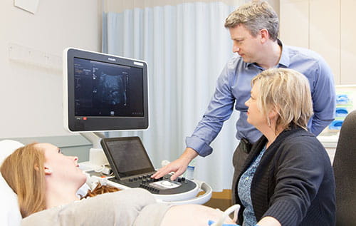 James Strainic, MD performing echocardiography.
By the mid-1990s, the association of increased nuchal translucency and fetal heart disease became apparent even in the absence of chromosomal anomalies. Nuchal translucency screening offered to all pregnant women is now the standard of care in many developed countries. Even in the setting of normal chromosome analysis, a nuchal translucency over the 95th percentile is associated with major CHDs. This risk increases with increasing nuchal translucency with a very high chance of CHD with a NT greater than 3.5mm.1 When early fetal echocardiography is inaccessible, women who receive a high-risk nuchal translucency result must endure a prolonged wait (typically over 2 months) until later fetal echocardiography is available.
In addition to those with increased nuchal translucency, several other well recognized high-risk indications for fetal CHDs are identifiable in early pregnancy:
- Abnormal ductus venosus flow
- Presence of tricuspid regurgitation
- Abnormal cardiac axis (normal 40-60 degrees)
- Presence of pericardial/pleural effusion
- Abnormal screens
- Maternal/paternal CHD or previous child with heart defect (especially when associated with high prevalence of gene disorders)
For those in whom an abnormality is detected, having time to process information on the nature of the condition, potential treatment options, anticipated outcomes and opportunities for genetic testing are invaluable. In addition, for those considering termination of pregnancy, there is less time pressure at this stage (well before the legal gestational age limits for termination of pregnancy in most states) and the procedure itself is safer and less psychologically traumatizing than later in pregnancy. In fact, at the time of early fetal echocardiography many women’s family and friends will be unaware of their pregnancy, resulting in fewer social ramifications of pregnancy termination. Many couples, however, do continue with the pregnancy and can use the extra time provided by early diagnosis to prepare and plan for a baby with additional health needs, including making practical and economical arrangements in order to minimize the disruption to their family lives.
Anxious parents with a child with congenital heart disease often experience post-traumatic stress in future pregnancies. “In our Fetal Heart Program and The Congenital Heart Collaborative, we really bond with families when they are going through all of the repairs or palliations for their children’s heart defects. Seeing their child in such a fragile state can be extremely anxiety producing. During a second pregnancy, our families often are frightened and nervous that they will have to go through that experience again. Offering reassurance at 11-14 weeks instead of 18-22 weeks can really alleviate that stress” says James Strainic, MD, Director of the Fetal Heart Program. They can receive valuable reassurance from a normal early fetal echocardiogram. The International Society for Ultrasound in Obstetrics and Gynecology (ISUOG) suggest that it is appropriate to offer a scan at or before 14 weeks’ gestation if the nuchal translucency is greater than or equal to 3.5mm, a mother is particularly anxious in view of a family history, or as soon as possible once congenital heart disease has been suspected (regardless of gestational age).
“Given that these conditions have a spectrum of severity and usually progress during gestation, it is critical to evaluate the fetus as early as possible,” says Ellie Ragsdale, MD, Maternal Fetal Medicine Specialist, UH Cleveland Medical Center and Assistant Professor of Obstetrics and Gynecology, Case Western Reserve University School of Medicine.
“Patients expect an uncomplicated pregnancy with a vaginal delivery at their community hospitals and a perfectly healthy baby,” she says. “They are so shocked when they learn their pregnancy is not going to take that course. With all the focus on what is being done for the baby, the mother can feel lost in the shuffle.”
She counsels women about how to manage comorbidities such as diabetes or hypertension, advises them on what to expect in any future pregnancies and works with genetic counselors to help women learn if the cardiac defect has a genetic cause.
Dr. Ragsdale notes that many institutions require women carrying a baby with a cardiac defect to deliver via Cesarean section, but the unique set up at UH Cleveland Medical Center, with a maternal hospital connected to a pediatric hospital, allows doctors to attempt a vaginal delivery in a traditional suite and be near the baby after birth. The collaboration between Maternal Fetal Medicine and the Fetal Heart Team provided under one roof ensures seamless care to patients throughout their pregnancy journey and beyond.
Doctors can refer a patient to one of our board-certified maternal fetal medicine specialists at UH MacDonald Women’s Hospital:
MFM Consultation | Call 216-844-8545
OB ultrasound | Call 216-844-7881
Genetic counseling | Call 216-844-3936
Fetal echocardiogram | Call 216-844-1337
The Fetal Heart Team
James Strainic, MD
Director, Fetal Heart Program
Assistant Professor
Case Western Reserve University School of Medicine
Sarah Plummer, MD
Assistant Professor
Case Western Reserve University School of Medicine
Tiffanie McCourt, MSN, APRN, CPNP AC-PC
Fetal Heart Program Coordinator
Rachel Leonard, RN
Contact the Fetal Heart Program to schedule a fetal echocardiogram and consultation: 216-844-1337
Maternal Fetal Medicine
David Hackney, MD
Division Chief, Maternal Fetal Medicine
Associate Professor
Case Western Reserve University School of Medicine
Jane Corteville, MD
Director, MacDonald Imaging
Associate Professor
Case Western Reserve University School of Medicine
Noam Lazebnik, MD
Professor
Case Western Reserve University School of Medicine
Melissa March, MD
Medical Director, Labor and Delivery
Assistant Professor
Case Western Reserve University School of Medicine
Ellie Ragsdale, MD
Director, Fetal Intervention
Assistant Professor
Case Western Reserve University School of Medicine
Elizabeth Ruzga, DNP, APRN-CNM
Assistant Clinical Professor
Case Western Reserve University School of Medicine
Christophe Nau, MD
Assistant Professor
Case Western Reserve University School of Medicine
Laura Petiya, NP
References
1. W Lee 1, L Allan, J S Carvalho, R Chaoui, J Copel, G Devore, K Hecher, H Munoz, T Nelson, D Paladini, S Yagel, ISUOG Fetal Echocardiography Task Force. (2008). ISUOG consensus statement: What constitutes a fetal echocardiogram? Ultrasound in Obstetrics & Gynecology : The Official Journal of the International Society of Ultrasound in Obstetrics and Gynecology, 32(2), 239-242. doi:10.1002/uog.6115 [doi]
Tags: Congenital Heart Disease